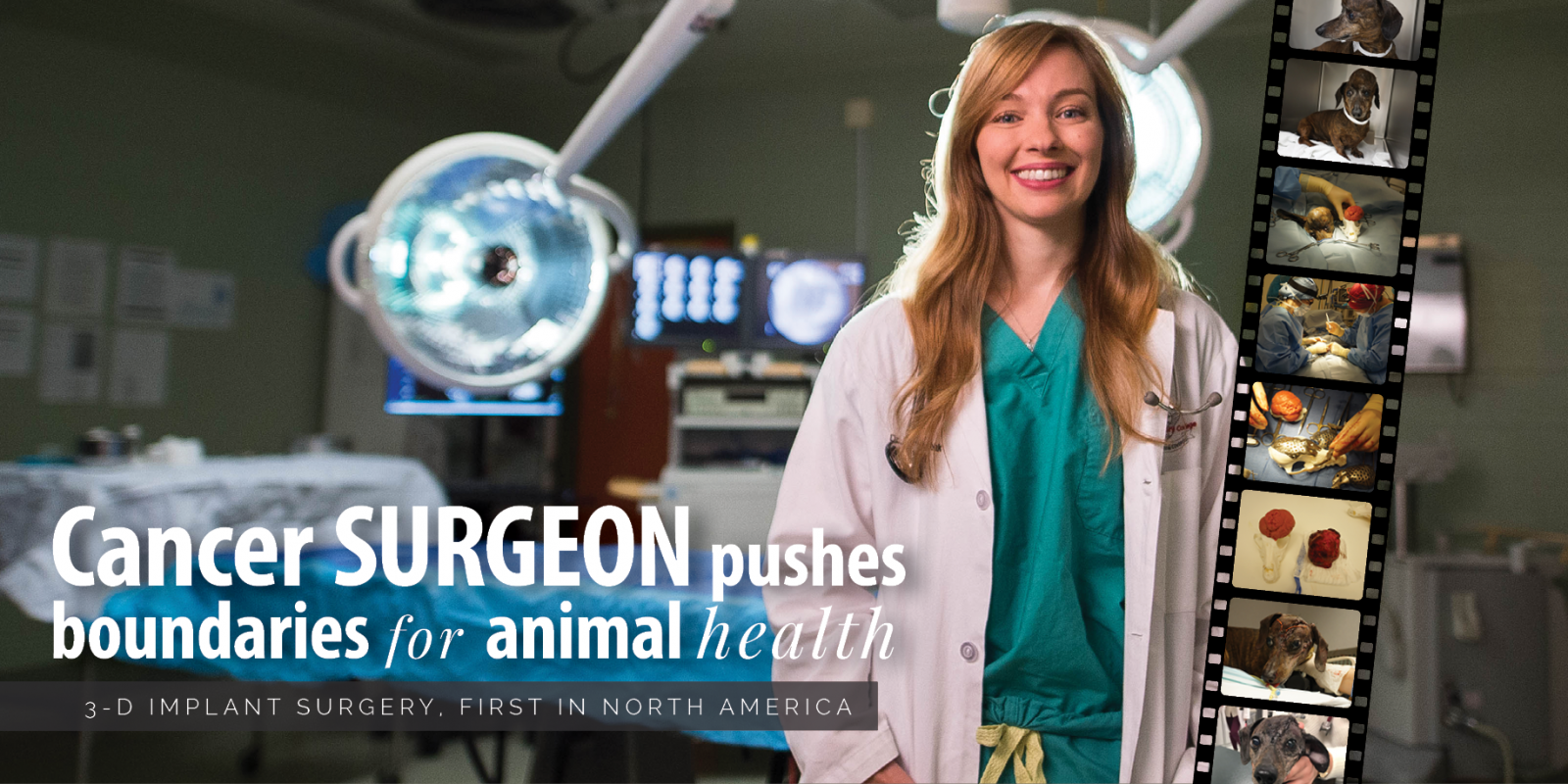
3-D implant surgery, first in North America
For the first known time in North America, Ontario Veterinary College (OVC) board-certified veterinary surgical oncologist Dr. Michelle Oblak led a successful reconstructive skull surgery implanting a custom 3-D printed skull plate in a dog.
When Patches, an eight-year-old dachshund, presented to Dr. Galina Hayes at Cornell University’s College of Veterinary Medicine with a large cancerous growth on her skull, Oblak’s former OVC colleague, and soft tissue surgeon, contacted her for advice on the difficult case.
As a veterinary surgical oncologist and an assistant professor in soft tissue surgery at the OVC, Oblak has a special interest in canine skull tumours and she recently published a book chapter on the topic. She has also been researching better ways to develop surgical plans and reconstruct the skulls in these patients. As part of this work, Oblak is a member of the OVC RaPPID (rapid prototyping of patient-specific implants for dogs) team. The group is looking at the feasibility of 3-D printed materials and rapid prototyping for surgical planning and patient-specific, personalized implants for dogs. Rapid prototyping involves constructing a model using 3-D computer-aided design data. When she heard about Patches and saw her CT scans, she thought that it might be the perfect case to offer this up-and-coming, cutting-edge surgical option.
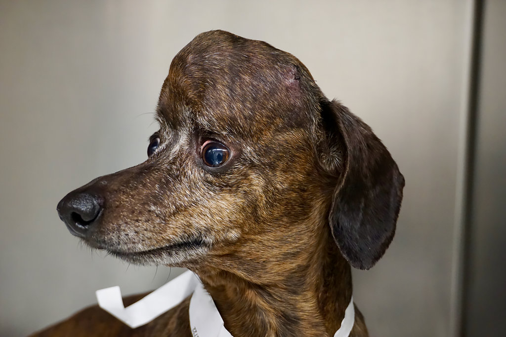
Since the cancerous tumour was growing within the bone of Patches’ skull, the surgical team estimated they needed to take out almost 75 per cent of the top area of her skull that covered her brain in order to remove as much cancer as possible.
“Given the complexity of the area and the amount of brain exposed, we knew we would need some sort of reconstruction after removing the tumour,” Oblak explains. “We also wanted to plan our surgical approach in advance, especially if we wanted to save Patches’ eye, which was being threatened by the tumour’s aggressive growth.”
Working with Dr. Alex zur Linden, a board-certified veterinary radiologist at OVC, and John Phillips, an engineer with Sheridan CAMDT, Oblak imaged and built a 3-D model of Patches’ skull. The team then worked in collaboration with ADEISS, a company that develops 3-D printed medical devices for human medicine, to access software designed for mapping and modelling human skulls for her dog patient. The software allowed her to plan the anticipated skull defect, as well as, design and print a custom titanium plate for Patches’ upcoming surgery. Less than a month after receiving Hayes’ phone call, with a physical skull model and personalized skull plate in hand, Oblak travelled to the United States to lend her expertise to the case.
Oblak and Hayes performed the three-hour surgery this past spring in New York. After removing the tumour, the implant was placed to protect Patches’ brain and soft skull tissue, and it fit perfectly. Oblak says without the technology, the team may have spent up to an additional two hours in the operating room, contouring a titanium mesh, an option that would not have had the same cosmetic outcome for Patches as the custom 3-D implant.
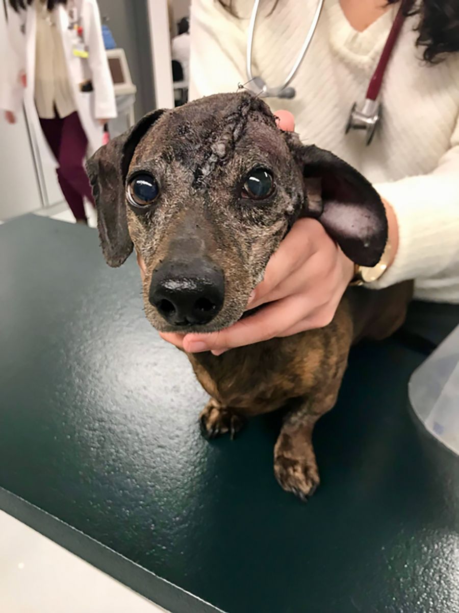
The surgery was an overwhelming success. Patches woke up normally from general anesthesia following the procedure and was alert and looking around within a half an hour. Oblak was able to offer a customized, state-of-the-art 3-D printed implant option to a canine patient and their family for the very first time in North America.
“I am thrilled I had the opportunity to provide something as ground-breaking as this procedure for a dog,” Oblak says. “The benefits of incorporating 3-D print technology into veterinary medicine are, and will continue to be, incredibly impactful – allowing us to offer faster, safer and improved surgeries for our pets. The benefits apply well beyond reconstructive surgery like Patches’. 3-D printed implants also have the ability to help in trauma, limb deformity and fracture cases as well,” she explains, adding that there is remarkable potential for rapid prototyping to help both humans and animals alike.
Oblak says one of the best parts of working in academia is to explore new, different and better ways to help her patients. “Innovation and progress is what inspires me each and every day.”
Dr. Michelle Oblak is an assistant professor and board-certified veterinary surgical oncologist at the Ontario Veterinary College at the University of Guelph. She is an OVC Pet Trust-funded researcher with a focus on translational medicine and has several collaborations considering dogs as a naturally occurring disease model for cancer in humans.
Feature photo: Dr. Michelle Oblak.
Photo reel, images from top to bottom:
Patches with her tumour, prior to surgery; Patches’ tumour with Oblak’s 3-D printed model, used to create the surgical plan; Drs. Oblak and Hayes conducting Patches’ surgery at Cornell University; Oblak shows how the titanium 3-D printed plate will fit on Patches’ skull with her 3-D model; The tumour, removed, right, with 3-D print model, left; Patches, wakes up after surgery; Patches has her stitches removed, two-weeks after her surgery.
Read more in the fall / winter issue of Best Friends Magazine.