 “The day I found out my dog had a rare type of cancer, I was completely terrified: I was shocked and I was devastated.”
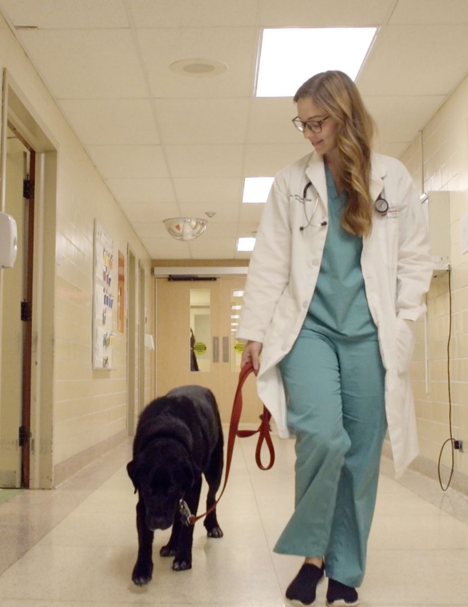
Kris Depowski rescued her beloved mixed breed dog, Murphy, in 2013 when he was one year old. He had been found emaciated, running down a highway in Tennessee. Kris heard about him through a rescue group and it was love at first sight, she remembers. It was devastating when Murphy’s family veterinarian in Buffalo, New York found a small mass the size of a golf ball on his skull earlier this year and recommended that Kris and her dog immediately travel to the University of Guelph’s Ontario Veterinary College (OVC) for further evaluation.
Murphy was diagnosed with multilobular osteochondrosarcoma (MLO), a type of cancer that starts in the bone. MLO tumours most commonly develop on flat bones such as the skull, as in Murphy’s case. These tumours are usually considered slow-growing but can become quite serious quickly given their location and proximity to the brain. Clinical signs in dogs may include a noticeable bump on the head and neurologic deficits, like seizures. MLOs can affect any size or breed of dog. Treatment requires complex surgical intervention and often the removal of the portion of the skull that is affected.
After Murphy’s initial consultation at the Mona Campbell Centre for Animal Cancer, the oncology service within the OVC Health Sciences Centre, Kris returned home to Buffalo with a tough decision to make on her dog’s behalf.
“I was feeling unsure. The decision I had to make was huge. I didn’t know enough about his cancer or potential outcomes since it was so rare, and so little research has been done on it. I pulled out my computer and started to investigate; it was then that I came across a story that had aired on CBS Evening News only a few months earlier. This same surgery that my Murphy needed had been done once before – in a dog with the same type of cancer as mine,” she remembers. “I watched OVC’s Dr. Michelle Oblak interview about the procedure and successfully replacing 70 per cent of a dog’s skull with a 3-D printed plate in another American dog, named Patches. It was a medical milestone and that was what turned the tide for me.”
Kris decided to move forward with Murphy’s procedure.
“The fact that this innovative treatment was even available to us, even though we had to travel to another country, was truly life-saving,” she says, understandably emotional. “As a dog mom, I was thrown into a cascade of emotion. It was a leap of faith. This was it. This was going to be Murphy’s chance to live.”
 Within a month, Kris returned to OVC, this time for a CT scan, and images of Murphy’s skull were taken. 3-D printing is instrumental in planning and performing surgeries such as Murphy’s.
“We used the CT scan to build a 3-D model of Murphy’s tumour and skull. Using the model, we can determine the best surgical path and even practice the complex surgery well in advance of being in the operating room (OR),” says Oblak. “On top of planning, the model allows us to reassure pet owners by demonstrating what the procedure will look like, show how the 3-D plate (also created from the scans) will be placed once the tumour is removed and answer any pressing questions or concerns they may have.”
.jpeg)
Oblak says the innovative technology arms her with the tools she needs as a veterinary surgical oncologist to make the major procedure a success before she even steps into the OR.
“After performing Murphy’s CT scan we found that his tumour was large and extended deep down into his sinus. Having this information and being able to plan everything ahead of time was crucial for the procedure’s success,” Oblak explains. “Since we could see that the tumour was behind his eye, it would have been difficult to create a traditional hand molded mesh implant that contoured well in that region. 3-D printing technology has allowed us to bring creative innovation to the clinic floor and directly helps us save lives in the OR.”
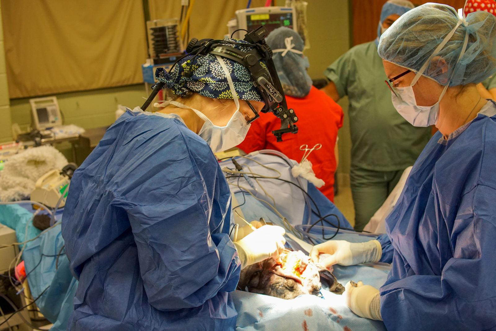
Murphy underwent a partial craniectomy on May 7, 2019 at the OVC Companion Animal Hospital.
“After the tumour was removed, the sterile implant was then carefully placed over the left side of Murphy’s skull. The ability to practice on a model was invaluable in our ability to save Murphy’s left eye,” Oblak explains.
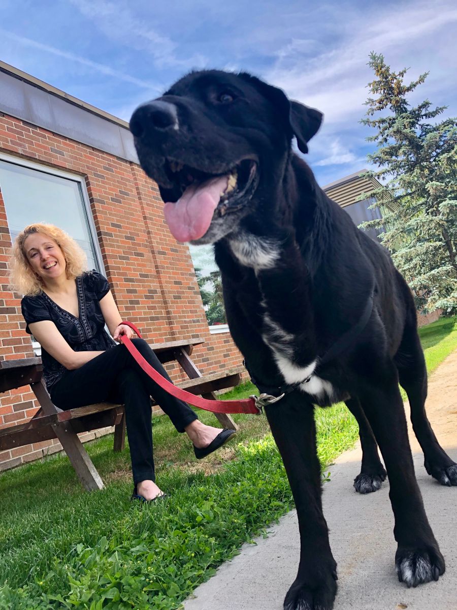
Murphy spent six days recovering in the OVC Intensive Care Unit after his surgery and was able to return home in May of this year.
The decision to bring Murphy to Canada was an easy one. Kris says that when she adopted him, she promised she would give her dog the best life possible. This surgery was the only option to save his life and it was only available at OVC.
“Dr. Oblak’s skill, innovative expertise and incredible compassion saved Murphy’s life. You can’t put a price on that,” Kris says. “The gratitude I feel is truly overwhelming. I am so thankful for the staff and doctors – it is because of OVC that I have my dog back. I owe them everything.”
Watch Murphy's story.
Read more in the fall / winter issue of Best Friends Magazine.